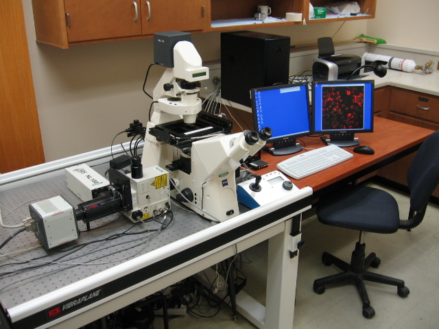
Spinning disk confocal microscope
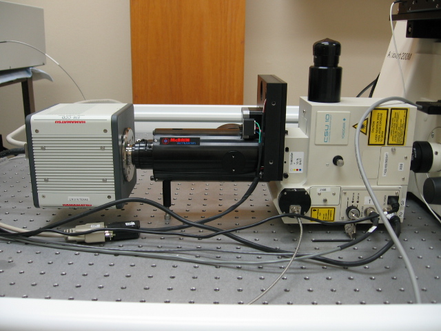
The centerpiece is a Zeiss 200M inverted optical microscope that has: 5 objectives, magnification power from 5 times to 160 times. Our 63x objective has a maximum xy resolution at 488 nm of 200 nm, a z resolution of 490 nm, and a depth of field of 1440 nm. There is a Piezo stage with a z axis resolution of 1.5 nm and a xy resolution of 50 nm. It also contains a reflector turret with slots for 5 cubes. There is an epi-fluorescence system attached that has an ultraviolet, a blue, and a green excitation filter. There is a SPOT camera with a 6.45 micrometer pixel size that can be used for bright field, DIC, and epi-florescent imaging. McBain Instruments prepared a confocal attachment that includes a CSU-10 Yokogawa confocal scanner, an emission filter wheel, four lasers, a Hamamatsu camera with an 8 micrometer pixel size, and associated electronics for hardware control. The four lasers provide the following excitation wavelengths: 405, 457, 488, 514, 532, 568, and 647 nm. The emission filter wheel contains the following filters (Center/FWHM): 450/35, 485/30, 520/35, 545/30,585/40, 593/45, 624/40, 692/40, and 810/50 nm.
We have the ability to perform ablation using a MicroPoint system from Photonic Instruments.
The CSU-10 scanning system can produce optical sections that have a depth of field as small as 0.88 micrometers, when using the 1.6 optovar. It can take an image in as low as 3 ms.
This entire system is controlled with the SimplePCI software from Hamamatsu Corporation. We also have the Imaris software package from Bitplane that is optimized for the construction of three-dimensional objects from z-scan data produced with a confocal microscope.