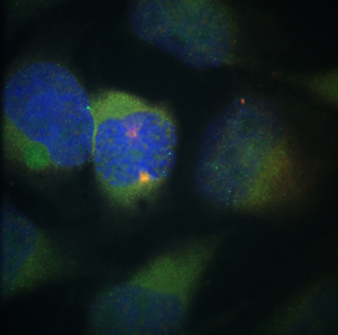
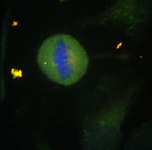
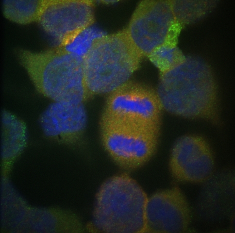
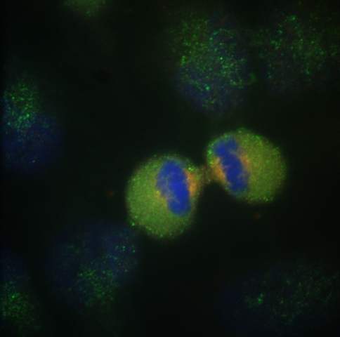
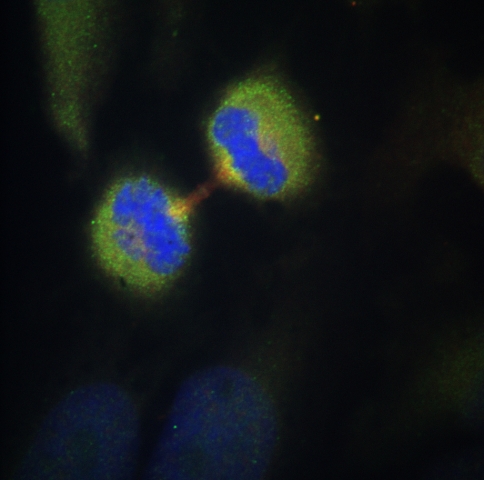
This is a set of images of cells undergoing mitosis: prophase, metaphase, anaphase, teleophase, and cytokineses.
The fluorophores that were used are: blue: Hoechst, Green: Fluorescein,
and Red: Texas Red. The fluorophores were excited with 405,
488 and 568 nm lasers. The images were collected with a Hamamatsu
camera set for a 64 gain for the blue channel and a 128 gain in the
green and red channels. The emission light was collected with
an 100 times magnification oil emmersion objective.
Sample prepared by Hemangi Patil, January 2009.